Symptoms
About Rotator Cuff Injury
Causes of Rotator Cuff Injury:
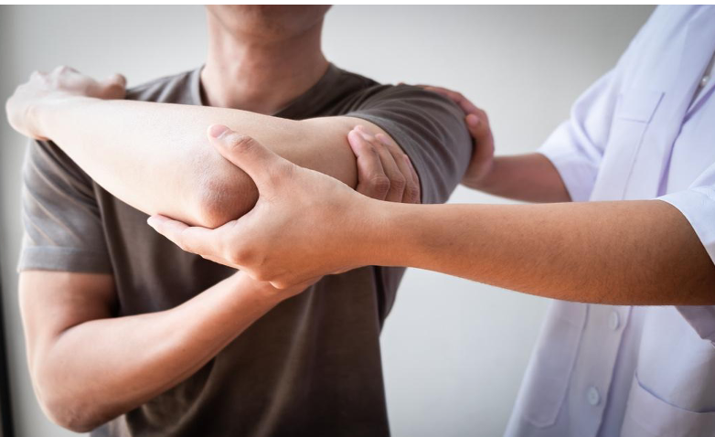
Rotator cuff injuries occur due to aging, overuse of the shoulder, or trauma. Problems with the shoulder bones or scapula, along with age-related degeneration of the tendons, can cause partial or complete tears in the rotator cuff.
Diagnostic Methods:
Diagnosis of a rotator cuff injury involves checking symptoms, testing shoulder movement and strength, and using imaging tests like X-rays or MRIs. These results are combined to determine the appropriate treatment plan.
Treatment through Physical Therapy:
If surgery is not performed, treatment focuses on pain relief, improving shoulder movement, strengthening muscles, and practicing daily activities. After surgery, the rehabilitation program starts with protecting the shoulder, gradually increasing movement, and rebuilding strength.
Prognosis:
The outcome of treatment varies from person to person, but many experience improved shoulder movement and reduced pain. Continued physical therapy is important to maintain shoulder function and prevent recurrence.

References
Burbank, K. M., Stevenson, J. H., Czarnecki, G. R., & Dorfman, J. (2008). Chronic shoulder pain: Part I. Evaluation and diagnosis. American Family Physician, 77(4), 453-460.
Chard, M. D., & Hazleman, B. L. (1993). Shoulder disorders in the elderly: a community survey. Rheumatology International, 13(3), 105-107.
Kuhn, J. E. (2009). Exercise in the treatment of rotator cuff impingement: A systematic review and a synthesized evidence-based rehabilitation protocol. Journal of Shoulder and Elbow Surgery, 18(1), 138-160.
Manske, R. C., & Prohaska, D. (2008). Postoperative rotator cuff rehabilitation: a literature-based rehabilitation protocol. International Journal of Sports Physical Therapy, 3(4), 265-281.
Yamaguchi, K., Ditsios, K., Middleton, W. D., Hildebolt, C. F., Galatz, L. M., & Teefey, S. A. (2006). The demographic and morphological features of rotator cuff disease: a comparison of asymptomatic and symptomatic shoulders. The Journal of Bone and Joint Surgery. American Volume, 88(8), 1699-1704.
About Sciatica
① Mechanism of Occurrence
Sciatica occurs when the sciatic nerve is compressed or inflamed. Common causes include herniated discs, which compress the nerve. Other causes may include spinal stenosis, bone spur formation, trauma, tumors, and the hardening of gluteal muscles.
② Physical Therapy Evaluation
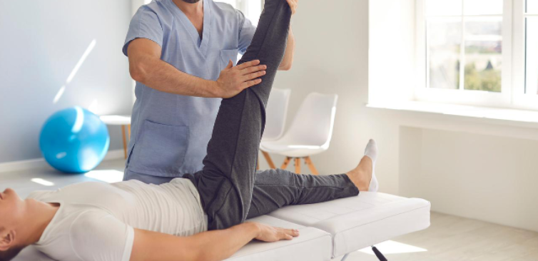
In physical therapy evaluations, posture and gait observation, muscle strength testing, range of motion measurement, and pain assessment are conducted. Specific neurological tests, such as:
– SLR Test (Straight Leg Raise Test)
– FNS Test (Femoral Nerve Stretch Test)
These are also performed to confirm the involvement of the sciatic nerve.
③ Physical Therapy Treatment
Physical therapy treatments include modalities such as heat therapy and electrotherapy to alleviate pain, exercises aimed at strengthening muscles and improving flexibility, and education on proper posture and movement patterns. Additionally, massage may be performed in some cases.
④ Prognosis of Sciatica
The prognosis of sciatica varies depending on the cause and the appropriateness of the treatment. In mild cases, natural recovery often occurs within a few weeks to months. However, severe cases may become chronic and require long-term management. Proper physical therapy and lifestyle improvements are crucial factors in ensuring a favorable prognosis.

References
1. van Tulder, M. W., Koes, B., & Bombardier, C. (2002). Low back pain. *Best Practice & Research Clinical Rheumatology*, 16(5), 761-775.
2. Fritz, J. M., & George, S. Z. (2000). The use of a classification approach to identify subgroups of patients with acute low back pain: Interrater reliability and short-term treatment outcomes. *Spine*, 25(1), 106-114.
3. Dagenais, S., Tricco, A. C., & Haldeman, S. (2010). Synthesis of recommendations for the assessment and management of low back pain from recent clinical practice guidelines. *The Spine Journal*, 10(6), 514-529.
About Low Back Pain
① Causes
Low back pain can be caused by various factors, including excessive strain on muscles and ligaments, disc damage, or joint issues. Having poor posture for overextended periods or sudden movements often lead to stress on the muscles and soft tissues in the lower back.
② Physical Therapy Evaluation
A physical therapist will observe the location of the pain and how symptoms change with different movements. They will also assess posture, flexibility, and muscle strength to identify the underlying causes of the problem. Specific tests involving certain movements are conducted to pinpoint the exact areas of concern.
③ Physical Therapy Treatment
Treatment typically involves heat or cold therapy to alleviate pain, exercises to strengthen the muscles around the lower back, and stretches to improve flexibility. Additionally, patients are educated on proper posture and movement patterns to prevent recurrence.
④ Prognosis
The prognosis for low back pain varies depending on the cause and treatment method. Mild cases often improve within a few weeks, while chronic low back pain may require long-term treatment. Proper physical therapy can accelerate recovery and reduce the risk of recurrence.

References
1. van Tulder, M. W., Koes, B., & Bombardier, C. (2002). Low back pain. *Best Practice & Research Clinical Rheumatology*, 16(5), 761-775.
2. Fritz, J. M., & George, S. Z. (2000). The use of a classification approach to identify subgroups of patients with acute low back pain: Interrater reliability and short-term treatment outcomes. *Spine*, 25(1), 106-114. 3. Dagenais, S., Tricco, A. C., & Haldeman, S. (2010). Synthesis of recommendations for the assessment and management of low back pain from recent clinical practice guidelines. *The Spine Journal*, 10(6), 514-529
About Meniscus Injuries
① Mechanism of Occurrence
Meniscus injuries occur when the knee is subjected to strong twisting or compressive forces. They are commonly seen during sudden changes in direction in sports or when the knee is hyperextended. As we age, the meniscus becomes weaker and more susceptible to injury.
② Physical Therapy Evaluation
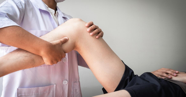
Physical therapists check the knee’s movement, pain, and swelling. They also perform tests to determine if there is a meniscus injury, such as:
– McMurray Test
– Apley Compression Test
③ Physical Therapy Treatment
Treatment includes cold therapy to reduce pain and inflammation, as well as exercises to strengthen the muscles around the knee. Depending on the severity of the injury, stretching exercises to improve knee flexibility may also be included.
④ Prognosis
The recovery from a meniscus injury depends on the extent of the damage and the effectiveness of the treatment. Minor injuries often heal within a few weeks to months, but severe cases may require surgery. Early physical therapy is key to improving the outcome.

References
1. Brophy, R. H., & Matava, M. J. (2010). Meniscal Injury. *Journal of the American Academy of Orthopaedic Surgeons*, 18(8), 527-536.
2. Englund, M., Guermazi, A., Gale, D., Hunter, D. J., Aliabadi, P., & Felson, D. T. (2008). Incidental Meniscal Findings on Knee MRI in Middle-aged and Elderly Persons. *New England Journal of Medicine*, 359(11), 1108-1115.
3. Papalia, R., Del Buono, A., Osti, L., Denaro, V., & Maffulli, N. (2011). Meniscectomy as a risk factor for knee osteoarthritis: A systematic review. *British Journal of Sports Medicine*, 45(8), 726-730.
About Flat Feet
① Causes
Flat feet occurs when the arches of the feet collapse, causing the entire foot to make contact with the ground. This condition can be caused by congenital factors, muscle weakness, loose ligaments, or excessive strain. While flat feet in children may correct naturally, in adults, they can lead to pain and fatigue.
② Physical Therapy Evaluation
A physical therapist will assess the height of the foot arch, walking patterns, foot and ankle movement, and muscle strength. By observing how the feet move while standing and walking, the therapist can determine how flat feet affects gait and posture.
③ Physical Therapy Treatment
Treatment typically includes the use of special insoles or shoes designed to support the foot arch. Exercises to strengthen the arch, along with stretches for the ankle and calf muscles, are also important. Learning proper walking and posture techniques is crucial to managing the condition.
④ Prognosis
The prognosis for flat feet depends on the cause and the type of treatment received. With appropriate treatment, symptoms can be reduced, and quality of life can be improved. Early intervention is particularly effective in minimizing pain and fatigue in daily activities.

References
1. Kirby, K. A. (2000). Biomechanics of the normal and abnormal foot. *Journal of the American Podiatric Medical Association*, 90(1), 30-34.
2. Kulcu, D. G., & Yavuzer, G. (2008). Flat foot and related factors in primary school children: a report of a screening study. *Rheumatology International*, 28(10), 1063-1068.
3. Kulig, K., Fietzer, A. L., & Popovich Jr, J. M. (2011). Ground reaction forces and knee mechanics in the weight acceptance phase of gait in women with excessive pronation. *Clinical Biomechanics*, 26(8), 823-828.
About Shin Splints
① Why They Occur
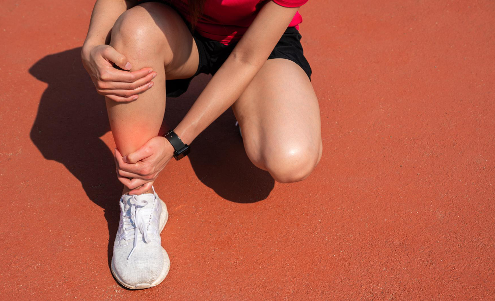
Shin splints are a condition that causes pain along the inner part of the shinbone. This happens when too much stress is placed on the shinbone and surrounding muscles, often due to high-impact activities like running or jumping. The shape of your feet can also make you more prone to developing shin splints.
② How They Are Evaluated
A physical therapist will assess where the pain is located and how your symptoms change when you walk or run. They will also examine the alignment of your feet, the flexibility, and the strength of your muscles, and analyze how you move.
③ How They Are Treated
Treatment usually involves resting and applying ice to reduce pain and inflammation. Stretching and strengthening the muscles around the shin are also important. Wearing the right shoes and using insoles can help prevent the condition from coming back.
④ How Long It Takes to Heal
With proper treatment and rest, shin splints typically heal within a few weeks to a few months. However, if left untreated, the symptoms can persist or even lead to stress fractures, so it’s important to address the issue early.

References
1. Michael, R. H., & Holder, L. E. (1985). The soleus syndrome. A cause of medial tibial stress (shin splints). *The American Journal of Sports Medicine*, 13(2), 87-94.
2. Thacker, S. B., Gilchrist, J., Stroup, D. F., & Kimsey, C. D. (2002). The prevention of shin splints in sports: a systematic review of literature. *Medicine and Science in Sports and Exercise*, 34(1), 32-40.
3. Craig, D. I. (2008). Medial tibial stress syndrome: evidence-based prevention. *Journal of Athletic Training*, 43(3), 316-318.
